
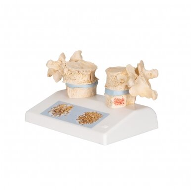
Osteoporozės didaktinis modelis
Kodas: 3B Scientific 1000182Kaina 10043 € su PVMPrekę turime sandėlyje. Užsakius ir apmokėjus šiandien iki 14 val., pristatytume kitą darbo dieną.
Šiuo metu prekės nėra sandėlyje. Numatomas pristatymo laikas nurodytas žemiau.
Pateikus užsakymą, susisieksime su Jumis ir informuosime apie tikslų numatomą užsakymo pristatymo laiką.
Šiuo metu prekės nėra sandėlyje. Numatomas pristatymo laikas nurodytas žemiau.
Pateikus užsakymą, susisieksime su Jumis ir informuosime apie tikslų numatomą užsakymo pristatymo laiką.
GamintojasPristatymo laikas3-6 savaitės-
Informacija apie prekę Prekės kodas 1000182 [A95] Svoris 0.238 kg Išmatavimai 14 x 9 x 10 cm Ženklas 3B Scientific
Osteoporosis Didactic Model - 3B Smart AnatomyNew anatomy app called 3B Smart Anatomy now included for FREE with Osteoporosis Didactic Model.
Every original 3B Scientific anatomy model now includes these additional FREE features:
- Free access to the anatomy course 3B Smart Anatomy, hosted inside the award-winning Complete Anatomy app by 3D4Medical
- The 3B Smart Anatomy course includes 23 digital anatomy lectures, 117 different virtual anatomy models and 39 anatomy quizzes to test your knowledge
- Bonus: FREE warranty upgrade from 3 to 5 years with every product registration
To unlock these benefits, simply scan the label located on your model and register online. All 3B Smart Anatomy features are completely free of charge for you.
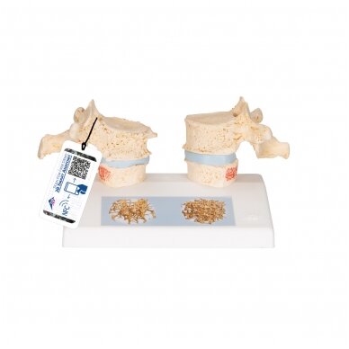
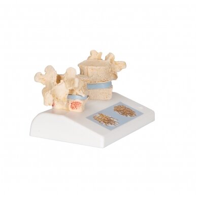
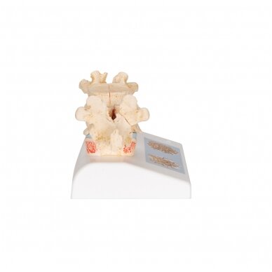
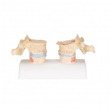
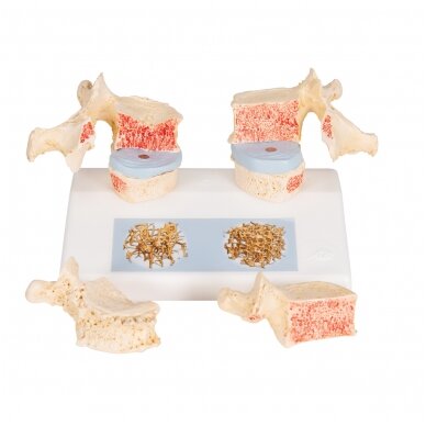
Impressive didactic model for comparing osteoporotic and normal thoracic vertebrae. Ideal for medical studies and patient consultation. The 11th and 12th thoracic vertebrae are shown.
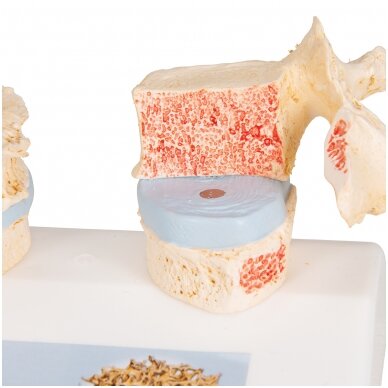
Reproductions of sequential osteoporotic thoracic vertebrae with narrower intervertebral disc are located on the left of the stand. The upper vertebra is divided in the middle. The magnetically attached vertebral half can be removed easily to show the cut surfaces. This allows clear visualization of the fractured upper part of the vertebral body caused by sintering, i.e. collapse of the bony substance in the course and as a result of osteoporosis. Degenerative changes in the bone, manifested as osteophytes, are also identifiable.
For comparison, reproductions of two corresponding healthy vertebrae with intervertebral disc are provided on the right side. One half of the upper vertebral body is magnetically attached and can be removed.
A detail illustration on the base depicts two 3D micro CT images obtained from bone biopsies. These illustrate the microacrchitecture of the osteoporotic bone, which has a lower bone density compared to healthy bone.
3B Smart Anatomy explained in 90 seconds:
Prekių krepšelis
Krepšelis tuščias.


