Rankos kaulo modelis su elastiniais raisčiais
Rankos kaulo modelis su elastiniais raisčiais
Kodas: 3B Scientific 1013683Kaina 31678 € su PVMPrekę turime sandėlyje. Užsakius ir apmokėjus šiandien iki 14 val., pristatytume kitą darbo dieną.
Šiuo metu prekės nėra sandėlyje. Numatomas pristatymo laikas nurodytas žemiau.
Pateikus užsakymą, susisieksime su Jumis ir informuosime apie tikslų numatomą užsakymo pristatymo laiką.
GamintojasPristatymo laikas3-6 savaitės-
Informacija apie prekę Prekės kodas 1013683 [M36] Svoris 0.24 kg Išmatavimai 14 x 10 x 28 cm Ženklas 3B Scientific
Hand Skeleton Model with Elastic Ligaments - 3B Smart AnatomyNew anatomy app called 3B Smart Anatomy now included for FREE with Hand Skeleton Model with Elastic Ligaments.
Every original 3B Scientific anatomy model now includes these additional FREE features:
- Free access to the anatomy course 3B Smart Anatomy, hosted inside the award-winning Complete Anatomy app by 3D4Medical
- The 3B Smart Anatomy course includes 23 digital anatomy lectures, 117 different virtual anatomy models and 39 anatomy quizzes to test your knowledge
- Bonus: FREE warranty upgrade from 3 to 5 years with every product registration
To unlock these benefits, simply scan the label located on your model and register online. All 3B Smart Anatomy features are completely free of charge for you.
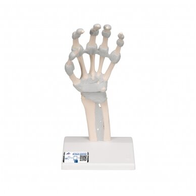
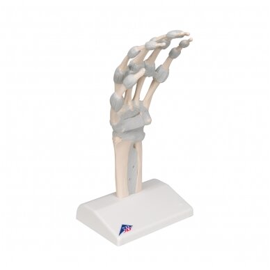
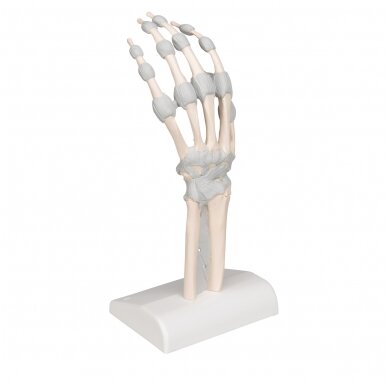
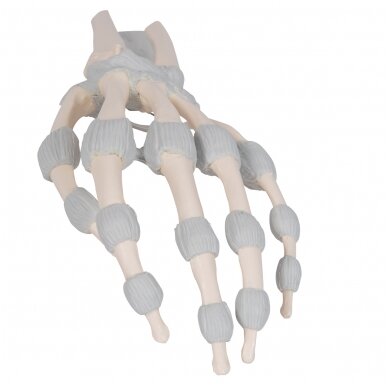
This single-part hand skeleton model shows the anatomy of the ligaments in the hand in detail. It is ideally suited both as a teaching aid as well as for anatomy classes, such as for medical students, physiotherapists and occupational therapists. The carpals (ossa carpi), the metacarpals (ossa metacarpi) and finger bones (ossa digitorum manus) are shown as osseous structures. In the distal area of the forearm, the radius and the ulna are represented.
- The fibrous layer of connective tissue, described in anatomy as the membrana interossea, is shown. It extends between both of these long bones.
- The retinaculum flexorum, which forms the top of the carpal tunnel, an anatomical area of clinical relevance, is also shown.
- All ligaments, the membrana interossea and the retinaculum flexorum are shown flexibly so that functional movements can be simulated for teaching purposes, in particular the joints in the wrist.
3B Smart Anatomy explained in 90 seconds:


